3D reverse engineering and simulation of signal flow in anatomically realistic neuronal networks
Marcel Oberlaender (Max Planck Florida Institute, Jupiter), Christiaan deKock (VU University Amsterdam), Randy Bruno (Columbia University, New York), Stefan Lang (IWR, Heidelberg), Bert Sakmann (Max Planck Florida Institute, Jupiter)
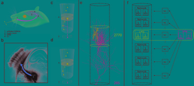
Soma location, dendrite morphology and presynaptic innervation represent key determinants of functional responses of individual neurons, such a sensory-evoked spiking. Here, we reverse engineer the three-dimensional networks formed by thalamocortical afferents from the lemniscal pathway and excitatory neurons of an anatomically defined cortical column in rat vibrissal cortex. We objectively classify nine cell types and quantify the number and distribution of their somata, dendrites and thalamocortical synapses. Somata and dendrites of most cell types intermingle, while thalamocortical connectivity depends strongly upon the cell type and the three-dimensional soma location of the postsynaptic neuron. Our dataset provides the first three-dimensional anatomical description of the cell type-specific lemniscal synaptic wiring diagram and elucidates structure-function relationships of this physiologically relevant pathway at single-cell resolution. Simulation of signal flow, evoked by passive whisker touch, revealed that the three-dimensional structure of thalamocortical networks in a cortical column can account for cell type- and location-specific subthreshold and spiking responses. Thus, the present approach may allow investigating the cellular and subcellular mechanisms that underlie whisker-mediated behaviors.


 Latest news for Neuroinformatics 2011
 Follow INCF on Twitter
