A realistic large-scale model of the retinal cone mosaic
Yoshimi Kamiyama (Information Science and Technology, Aichi Prefectural University), Daiki Sone (Information Science and Technology, Aichi Prefectural University), Shiro Usui (RIKEN Brain Science Institute)
The physiology and anatomy of the retina are relatively well known. Physiological studies of the retina have uncovered a number of cellular and subcellular mechanisms such as the characteristics of the ion channels found in retinal neurons. Anatomical studies have revealed the morphological principles hidden in the structure of the retina. It is as yet impossible to reconstruct a complete model of the retina. But it is now possible to identify some computational operations that the retina performs and to relate them to specific physiological and anatomical mechanisms, on the basis of neuroinformatics.
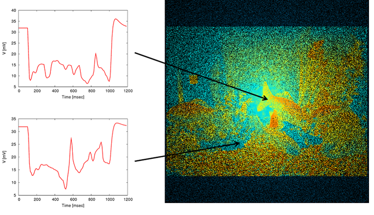
In the present study, a realistic model of the retinal cone mosaic is proposed. The spatial arrangement of the photoreceptors, i.e. photoreceptor mosaic, samples the continuous image of the world that is focused on the retina and transforms the image into a discrete array of signals that is transmitted to higher stages in the visual information processing. Color vision is mediated by three types of cone photoreceptors, the L, M and S cones. These cones have the function of converting absorbed photons into neural signals with different peak sensitivities at long (L), medium (M) and short (S) wavelengths. The cones distribute nonuniformly: cone density decreases with distance from the fovea, the L/M cone ratio are highly variable between individuals, and S cones are absent from the center of the fovea. We developed a large-scale computational model of the cone photoreceptors based on these physiological and anatomical characteristics. The model incorporated the spectral sensitivities of three types of cones, as well as the nonuniform spatial distributions of cones. The cone mosaic was generated by a stochastic algorithm to reproduce the nonuniformity. The chromatic light response was modeled by the spectral sensitivity and the ordinary differential equations of the membrane dynamics. The present model covers a visual angle of 60 degrees with two million cones. The model was implemented on the Riken Integrated Cluster of Clusters with 1024 nodes. In simulation, the model reproduced the nonuniform distribution of cones similar to the anotomical data. The model also well reproduced the chromatic light responses of the cones. In conclusion, the present model makes it possible to analyze how color information is processed in the retinal cone mosaic.


 Latest news for Neuroinformatics 2011
 Follow INCF on Twitter
