A toolbox for the visualization and metaanalysis of functional organization of the cortex using an anatomical database
Timothy J Herron (US Veterans Affairs Research), Anthony D Cate (US Veterans Affairs Research), Xiaojian Kang (UC Davis Neurology and Center for Neuroscience), David L Woods (US Veterans Affairs and UC Davis)
The MatLab toolbox VAMCA (Visualization And Meta-analysis on Cortical Anatomy) [www.nitrc.org/projects/vamca] provides 3D and surface-based meta-analyses of mean cortical functional activations that are published as stereotaxic 3D coordinates. VAMCA uses a database of cortices from 60 young, healthy, right-handed subjects to locate activations on a standardized cortical surface by extending the technique of multi-fiducial mapping [NeuroImage 28:635 2005] in order to perform meta-analyses. Non-parametric statistical tests are provided for determining (i) whether two groups of foci are in the same cortical location; (ii) the extent of overlap of the two groups’ foci; and (iii) whether two groups of foci are differentially concentrated in any of the anatomically defined regions of interest (ROI) as defined using FreeSurfer [surfer.nmr.mgh.harvard.edu].
First, we report on a new VAMCA email service that allows users to send stereotaxic coordinates in plain text or attachments to be processed and returned in minutes without any user setup or direct web access. The email-handling wrapper is relatively simple as well as moderately extensible and can leverage multiple computers to process email requests.
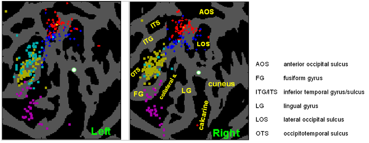
Second, we apply VAMCA to the non-trivial problem of localizing cortical activation coordinates to surface anatomy for the purposes of establishing functional ROIs useful for future fMRI group analyses on the cortical surface. As a sample analysis, we try to establish boundaries for several extrastriate visual stimulus selective regions [SSRs] based upon 3D coordinates from many visual localizer scans taken from hand-selected studies. We determine each SSR’s compactness and distinctness from/overlap with other similarly defined SSRs. We also use 55,000+ systematically collected coordinates from 6 journals within the SumsDB database [van Essen and Reid; sumsdb.wustl.edu/sums] to investigate how selective each SSR is across a wide gamut of functional contrasts.


 Latest news for Neuroinformatics 2011
 Follow INCF on Twitter
