EEG Source Estimation via Cortical Graph Smoothing
David Hammond (NeuroInformatics Center, University of Oregon), Benoit Scherrer (Computational Radiology Laboratory, Children's Hospital, Boston MA)
Introduction
The EEG source estimation problem consists of inferring brain activity from electrical potentials measured on the surface of the scalp. This is a challenging inverse problem as the number of degrees of freedom of activity inside the brain greatly exceeds the number of available measurements (e.g. scalp electrodes). Within the past ten years, the development of diffusion weighted imaging and associated modalities (e.g. DTI, DSI, HARDI, etc) has enabled non-invasive measurement of the anatomical connections between brain regions of individual subjects. In this work, we seek to exploit the availability of connectivity information in order to improve EEG source localization. We describe the cortical connectivity using a symmetric weighted graph, and introduce a prior for source activity by penalizing the sums of squares of differences of cortical activity across the edges. This encourages solutions which have consistent activation across connected cortical patches.
Methods
This work is done in the context of ongoing work on building accurate numerical models of human head electromagnetics, for EEG and ERP analysis [1]. Subject specific models are based on head tissue geometry segmented from a T1 MRI image, using the BrainK software package developed at the NeuroInformatics Center. The cortical surface is computed as the outer surface of segmented gray matter, and partitioned into Nd=2400 roughly equal area patches. Cortical sources are modeled by placing current dipoles at the center of each of the cortical patches, oriented perpendicular to the cortical surface. The relation between source currents J ∈ RNd and measured potentials Φ ∈ RNe at the electrodes, in the noise-free case, is given by Φ = KJ, where the columns of the Ne×Nd lead-field matrix K are determined by solving the inhomogeneous Poisson equation describing the electromagnetics of volume conduction.
We compute the connectome matrix by aligning the partitioned cortical surface with tract streamlines computed by whole-brain tractography, using software developed at the Computational Radiology Laboratory. The raw data for tractography consists of 10 unweighted (b=0) and 60 directional diffusion weighted images (b=700 s/mm^2), which are used to compute the diffusion tensor image. The tractography algorithm employed differs from simple streamlining through the use of both tensor deflection and directional inertia, which are employed to encourage tract streamlines to pass through regions of reduced anisotropy caused by crossing fibers. Additionally, sub-voxel resolution of the tract trajectory is enabled by use of log-Euclidean tensor interpolation. Tracts are initialized by seeding 30 random points in all image voxels with fractional anisotropy exceeding 0.4, yielding approximately 15 million streamlines which were then thresholded by retaining only tracts with both endpoints within 1 cm of the cortical surface. Start and end regions for each tract in this reduced set were determined from the cortical patches closest to the tract endpoints. The i,j entry of the Nd×Nd tractography-based connectome matrix Atr is then given by summing the reciprocals of tract lengths, for all tracts connecting regions i and j.
In addition to tractography, which captures the long-range non-local connections between cortical regions, we also include a local component of the connectome based purely on spatial adjacency of cortical patches. This local component is denoted Aloc, given by aloci,j=1 if cortical patches i and j are adjacent on the cortical surface, or 0 otherwise.
The overall weighted graph connectome has adjacency matrix A=τtr Atr + τloc Aloc, where τtr and τloc are regularization constants. The graph smoothing penalty is given by ∑i,jai,j(Ji −Jj)2.
This expression can be written as JT L J, using the graph Laplacian operator L defined by L=D-A, where D is the diagonal degree matrix with nonzero entries di,i=∑j ai,j
The cortical graph smoothing (CGS) estimate for the source currents, given observed potentials Phi at some timepoint, is defined as the solution to
Jgs = argminJ ||Φ−KJ||2+τtr JT Ltr J + τloc JTLloc JResults
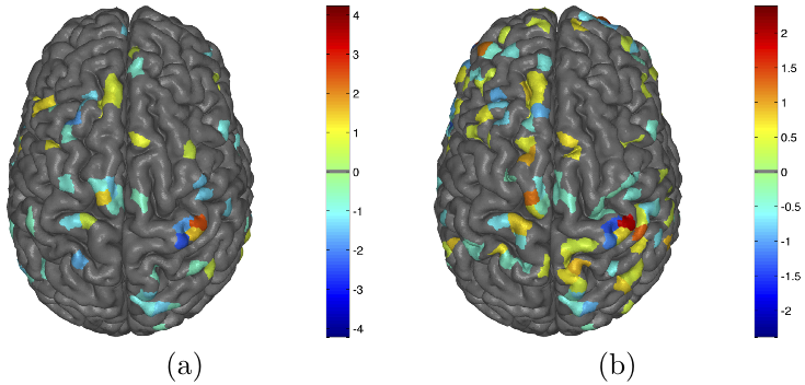
As a preliminary assessment of the utility of the CGS method, we show source estimation results for event related potential (ERP) data. In this motor potential study, collected at the NeuroInformatics Center and Electrical Geodesics Inc, subjects had high density (256 channel) EEG recorded while performing a button pushing task (left/right thumb/pinky) in response to a visual cue (color change of fixation point). ERP signal for each of the four digits (RP,LP,RT,LT) was generated by averaging a large number (>100) of trials, synchronized by time of the button push. Figure 1 shows both CGS and minimum norm source estimation results for a single subject, at 40 ms before left thumb button push. The part of the ERP signal related to the motor potential would be expected to be localized in the right precentral gyrus, in motor cortex known to be associated with the hand. While activity in this region is observed for both estimation methods, the CGS solution shows a sparser, more focal pattern of activity.
Conclusion
We have introduced a novel framework for regularization of cortical activity for EEG source localization, based on assuming smoothness on a cortical connectome graph computed using both white matter tractography and cortical surface adjacency. Additionally, we have demonstrated the new method yields sensible source estimates on ERP data for a simple motor task.
References
[1] A. Malony, A. Salman, S. Turovets, D. Tucker, V. Volkov, K. Li, J. Song, S. Biersdorff, C. Davey, C. Hoge, and D. Hammond, “Computational modeling of human head electromagnetics for source localization of milliscale brain dynamics,” Medicine Meets Virtual Reality, 2011.


 Latest news for Neuroinformatics 2011
 Follow INCF on Twitter
