Hemispheric asymmetries in the resting-sate functional connectivity patterns of the brain regions critical for language comprehension
And U. Turken (Veterans Affairs Northern California Health Care System, Martinez, CA), Nina F. Dronkers (Veterans Affairs Northern California Health Care System, Martinez, CA; and UC Davis Neurology Department, Davis, CA)
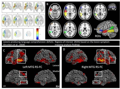
Introduction. We recently investigated the structural and functional connectivity profiles of five left hemisphere brain regions that support language comprehension (Turken & Dronkers, 2011). These regions were previously identified in a voxel-based lesion-symptom mapping (VLSM) investigation of auditory sentence comprehension deficits in aphasic patients with left hemisphere brain lesions due to stroke (Dronkers et al., 2004, Figure A). Lesion analysis findings highlighted as being critical for language comprehension the posterior middle temporal gyrus (MTG, yellow), anterior section of the superior temporal gyrus and Brodmann’s area 22 (ant. STG/BA22, red), Brodmann’s area 47 in the orbital section of the inferior frontal gyrus (IFGorb, BA47, blue), a part of Brodmann’s area 46 in middle frontal gyrus (BA46, brown) and a region extending from the superior temporal sulcus (STS) to parts of Brodmann’s area 39 (STS/BA39, green). Our subsequent analysis of structural and functional connectivity of these regions in a group of healthy subjects showed that these regions are part of a large network of brain regions that support language, interconnected by six major pathways and extending in the two hemispheres (Turken & Dronkers, 2011). The left posterior MTG showed a particularly extensive structural and functional connectivity pattern (Figure C), suggesting a key role in language comprehension. In order to gain further insights into the neural architecture of the language network, we examined here the hemispheric asymmetries in the functional connectivity patterns of these five regions.
Methods. Whole-brain functional connectivity patterns of each of the five regions and their right hemisphere homologues were assessed and compared between the two hemispheres using resting-state functional MRI data from 25 healthy subjects (1000 Functional Connectomes database, http://www.nitrc.org/projects/fcon_1000, NYU test-retest reliability dataset, Shehzad et al., 2009; 3T EPI, full cerebral coverage, 2 second TR, 6 minute runs in three separate session). The Functional Connectivity Toolbox (http://www.nitrc.org/projects/conn) for SPM8 was used for deriving the functional connectivity maps for each region of interest (ROI) defined in Montreal Neurological Institute (MNI) space. Hemispheric asymmetries were assessed using contrasts comparing at each voxel the strength of the coupling between that voxel and the left and right hemisphere homologues of each ROI. Group level analysis were conducted with voxel-wise t-tests (p < 0.05, Bonferroni correction, cluster extent > 25 voxels).
Results & Discussion. Hemispheric asymmetries in the resting-state functional connectivity patterns of the left and right posterior MTG were found for the angular gyrus, the orbital part of the inferior frontal gyrus (BA47), dorso-medial frontal cortex and the medial orbitofrontal cortex (Figure C, D). In each case, functional connectivity within the left hemisphere was stronger than in the right hemisphere. The anterior BA/BA22 did not show any significant asymmetries. The connectivity between BA47 and the angular gyrus as well as superior frontal gyrus was stronger in the left than in the right hemisphere. STS/BA39 showed stronger functional connectivity in the left hemisphere with IFGorb (BA 47) and dorso-medial frontal cortex. Overall, the results indicate stronger intrahemispheric coupling among the left hemisphere regions involved in language comprehension than between the homologous regions in the right hemisphere. The most notable functional connectivity asymmetries, involving the MTG, the angular gyrus and the orbital section of the IFG could be associated with structural asymmetries in the pathways connecting these regions, in particular, the middle longitudinal fasciculus and the inferior occipito-frontal fasciculus/extreme capsule fiber systems.
- Turken, A. U., Dronkers, N. F. (2011). The neural architecture of the language comprehension network: converging evidence from lesion and connectivity analyses. Frontiers in Systems Neuroscience, 5, 1:20.
- Dronkers, N.F., Wilkins, D.P., Van Valin, R.D., Jr., Redfern, B.B., Jaeger, J.J., 2004. Lesion analysis of the brain areas involved in language comprehension. Cognition 92, 145-177.
- Shehzad, Z., Kelly, A.M., Reiss, P.T., Gee, D.G., Gotimer, K., Uddin, L.Q., Lee, S.H., Margulies, D.S., Roy, A.K., Biswal, B.B., Petkova, E., Castellanos, F.X., Milham, M.P., 2009. The resting brain: unconstrained yet reliable. Cereb Cortex 19, 2209-2229.


 Latest news for Neuroinformatics 2011
 Follow INCF on Twitter
