Multicompartmental Model of Neurons Expressing Channelrhodopsin
Konstantin Nikolic (Imperial College London), Nir Grossman (Imperial College London)
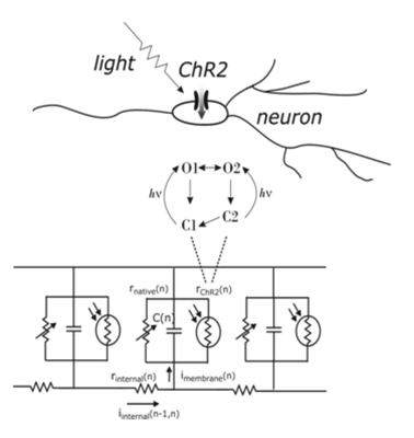
Optogenetic technology combines genetics and optics to create tools for controlling and interfering with cell functions. In the case of neuroscience it is a very important tool for interfacing with neurons and neural circuits. Light-sensitive proteins such as channelrhodopsin-2 (ChR2) or halorhodopsin (NpHR) are also ion channels or pumps and can be genetically expressed in the host cells. The specific parts of cells where expression occurs become light sensitive and external light sources can be used, in very precise spatio-temporal fashion if needed, to remotely generate ionic flux which causes membrane depolarisation or hyperpolarisation, depending on the type of ion channels. Design of various mutants of ChR2 and other light sensitive ion channels and pumps is now well within the reach of modern molecular biology. In contrary to conventional electrical stimulations, the ChR2 evoked currents are sensitive to the state of neurons they drive. ChR2 behaves as a complex light-controlled, voltage-dependent current driver (1-3), coupled to a dynamic-threshold voltage-oscillator (neuron). We developed a model of neurons expressing ChR2 which includes: (1) a photocycle model of ChR2 which gives the time evolution of functional states of ChR2 complex as a function of time and light intensity [1,2], (2) the current-voltage characteristic of ChR2 ion channels [3] (zero reverse potential was assumed, as suggested by experiments), (3) full mutlicompartmental Hodgkin-Huxley type model of the neuron [3]. The simulation code was developed on two platforms: Matlab and Simulink and it is available upon request. The model and the conclusions presented in this study can help to interpret experimental results, design illumination protocols and seek improvement strategies in the nascent optogenetics technology. The software is a very useful tool for computational modelling of networks of neurons which incorporate photosensitized neurons, as well as for simulations of neural computations within individual neurons.
Acknowledgment: European Commission, FP7-ICT-2009, Proj. No. 270324
[1] K. Nikolic et al. (2009). Photocycles of channelrhodopsin-2. Photochemistry & Photobiology 85, 400-411
[2] K. Nikolic et al. (2006). Modeling and engineering aspects of ChannelRhodopsin2 system for neural photostimulation, Proc 28th Conf IEEE Eng Med Biol Soc, 1626-1629
[3] N. Grossman et al. (2011). Modeling Study of the Light Stimulation of a Neuron Cell with Channelrhodopsin-2 Mutants. IEEE Trans Biomed Eng, in-press ISSN:1558-2531


 Latest news for Neuroinformatics 2011
 Follow INCF on Twitter
